What is magnetic resonance imaging (MRI)?

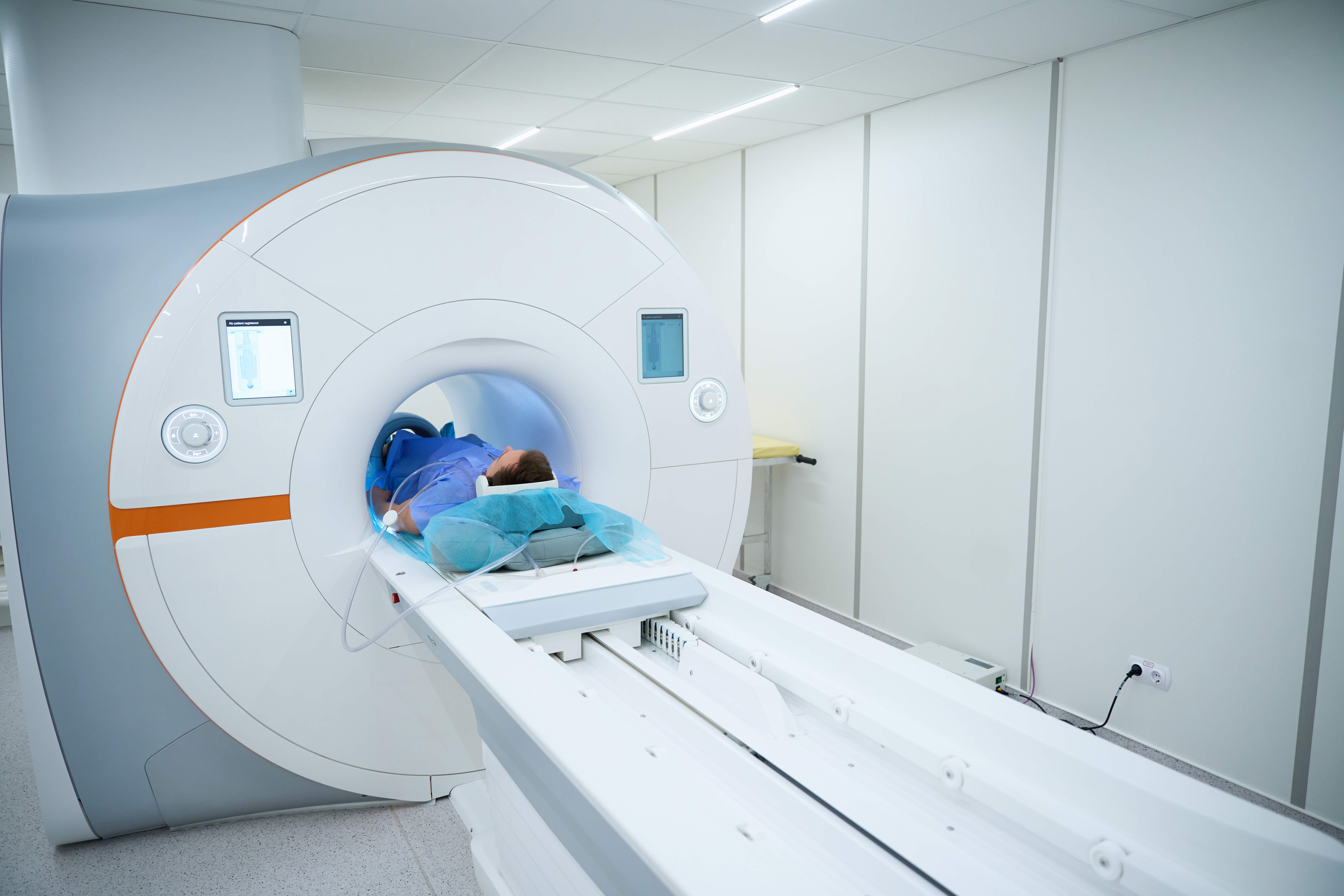
Magnetic resonance imaging (MRI) is a type of scan used to get a detailed picture of the inside of your body. 

  • It uses strong magnetism, radio waves and a computer
  • It does not use radiation (X-rays).

MRI cannot diagnose prostate cancer on its own. You will usually need to have a prostate biopsy as well. 


  • Watch this video from Cancer Research UK to find out what it is like to have an MRI scan.

© Cancer Research UK [2002] All rights reserved. Information taken 21/03/23. Cancer Research UK are independent from Prostate Cancer Research.

Important things to know

Why do I need an MRI scan?

Doctors use MRI scanners to look for any suspicious areas in:

  • Your prostate
  • The area surrounding your prostate. 

If they think that cancer may be present, they may recommend you have a prostate biopsy. 
Frequently asked questions (FAQs) about MRI scans
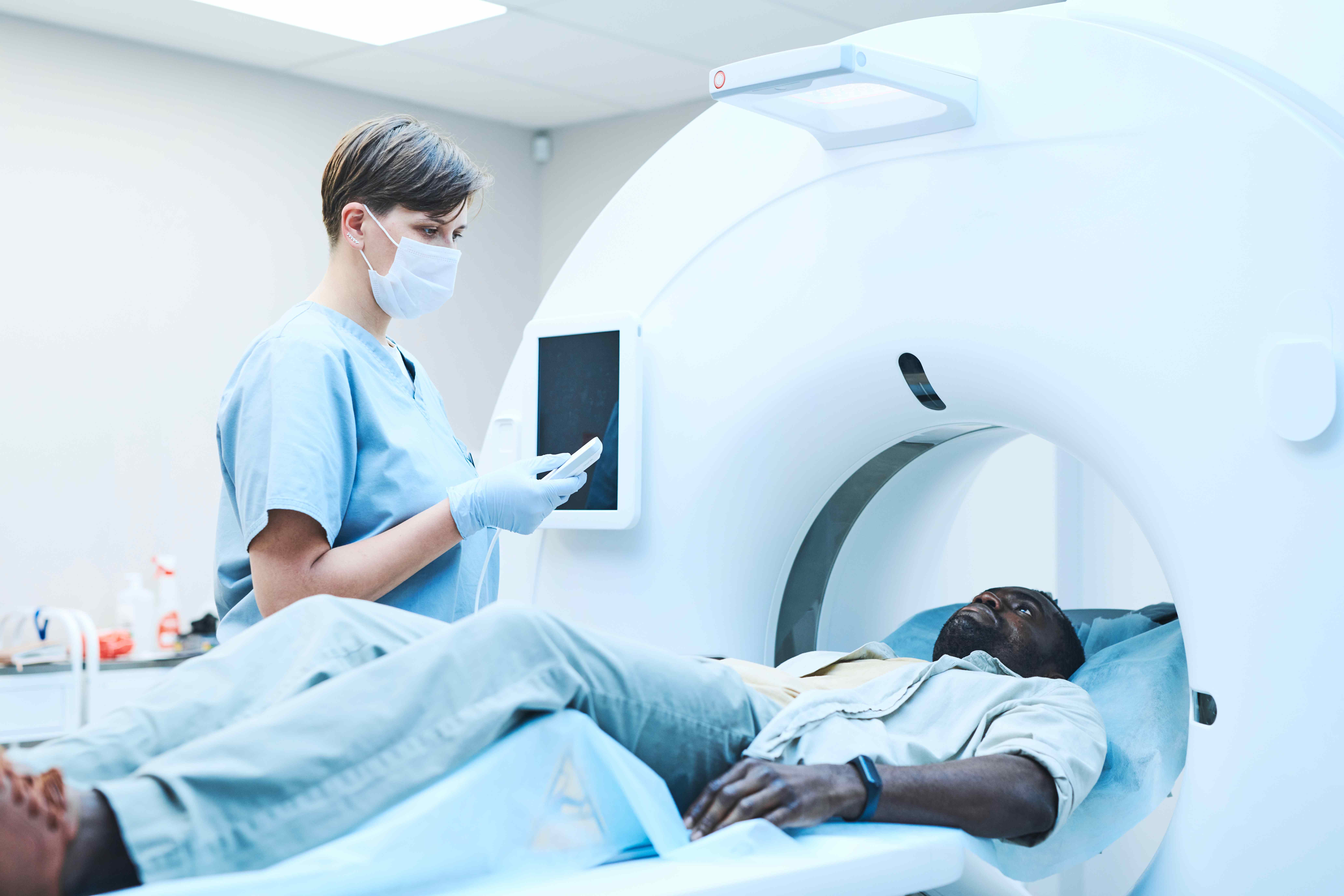
  • A standard MRI scanner is a large, cylinder-shaped tube. You will be asked to lie on a table that then slides into the tube. 


Black man in MRI scanner


  • Some people can feel shut in (claustrophobic) when inside the tube. If you think you may have a problem, contact the radiography department, or tell your doctor. Your doctor may prescribe you some light sedation. This will help you to relax. 


  • You will not be able to go into an ‘open’-type MRI scanner. This is because the magnetic field strength is lower than in closed MRIs. This means they will not give a clear enough picture of your prostate. 


  • MRI scans can be very loud. You may be given some ear plugs or headphones to wear before going into the scanner. 
  • A multi-parametric MRI (mpMRI) works by producing more detailed pictures of your prostate than a normal MRI. This means that your healthcare team can:
    • See any suspicious areas more easily.
    • Decide whether you need to have a biopsy of your prostate.
    • Target biopsy needles more accurately. This can reduce the number of samples that are taken from your prostate at one time.
    • See if there are any suspicious areas outside your prostate. For example, in lymph nodes or bones in your pelvis. 


  • It is often done before you have a prostate biopsy. 


  • mpMRI is now the ‘gold standard’ recommended type of MRI. But it is not available in every hospital. You can talk to your doctor about mpMRI. 


  • Our services search tool (link will open in a new tab) will allow you to search for hospitals near you that offer mpMRI.


What do I need to do?

  • You may be asked to empty your bowels before your MRI. Your appointment letter should tell you about any preparation you need. 


  • Some metals interfere with the magnetic field of the MRI. They may damage some of your belongings. Before going to the MRI room, you will need to remove:
    • Jewellery 
    • Watches, including electronic watches and GPS trackers
    • Mobile phones
    • Body piercings
    • Removable dental devices such as, braces
    • Pens
    • Spectacles
    • Credit cards
    • Hearing aids.


  • The radiographer will go through a safety checklist with you before you can have your MRI. You need to tell them about any metalwork that you have inside your body. This includes:
    • Pacemakers
    • Cochlea implants
    • Surgical clips, coils, pins or plates 
    • Dental fillings or bridges
    • Any other metal fragments.


  • Having any of these devices does not necessarily mean you will not be able to have an MRI. If you got a leaflet after having an implanted device, it may mention MRI. If you have these leaflets, bring them with you to the scan.

What is contrast medium?

  • You may have to have an injection of special dye. This is called a contrast medium. 
  • This will give a clearer picture of your prostate. 
  • You may also be given an injection that will stop bowel movement. This is called Buscopan (hyoscine butylbromide). 
  • Before you have the injections, tell the radiographer about any:
    • Allergies that you have
    • Medical conditions that you have.
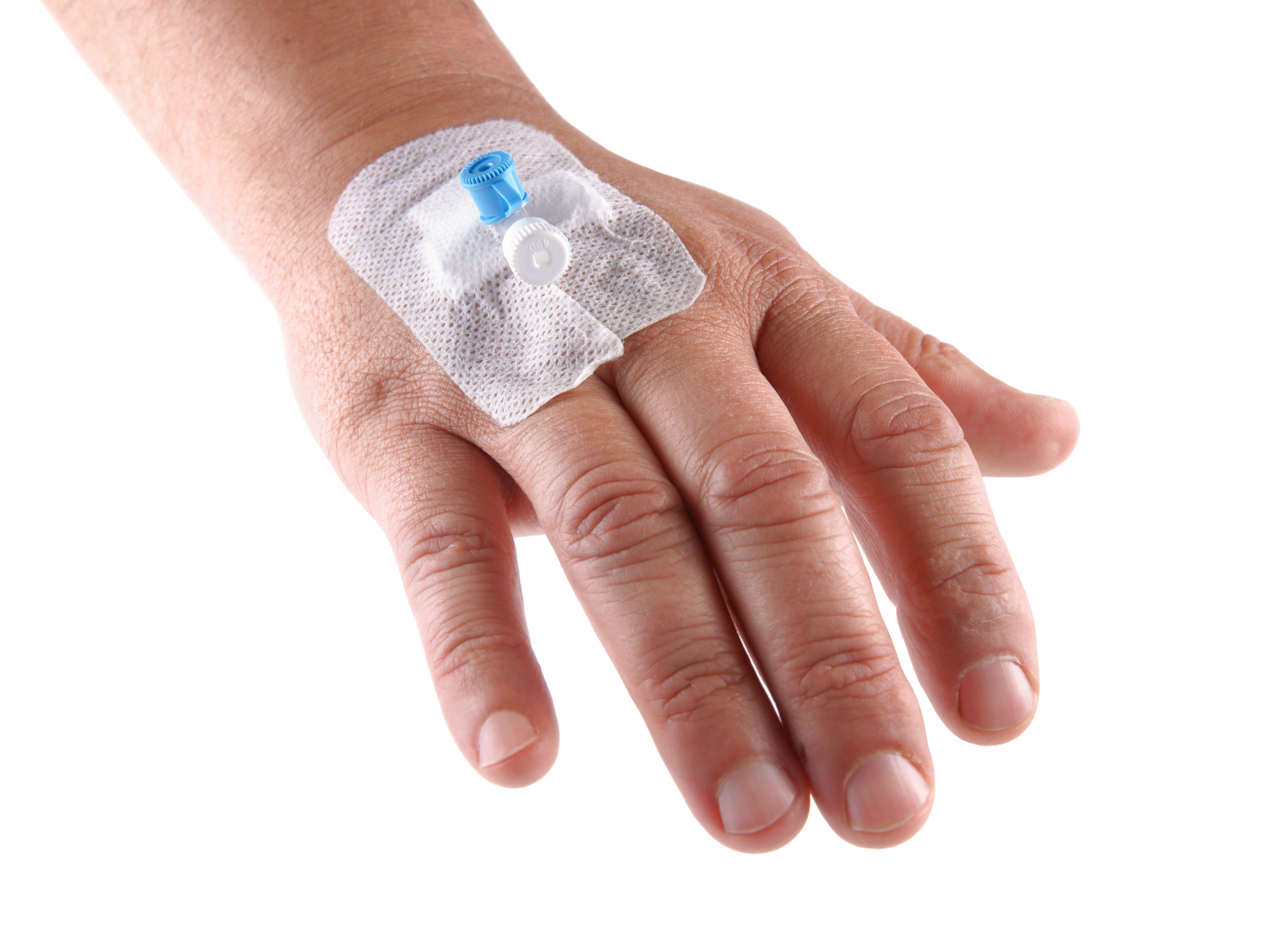
  • The radiographer will put a small tube into a vein on the back of your hand or in your arm. This is called a cannula. The radiographer willcannula in hand use this to inject the dye and the Buscopan. 
 

  • Your test results may take one-to-two weeks to come back. 


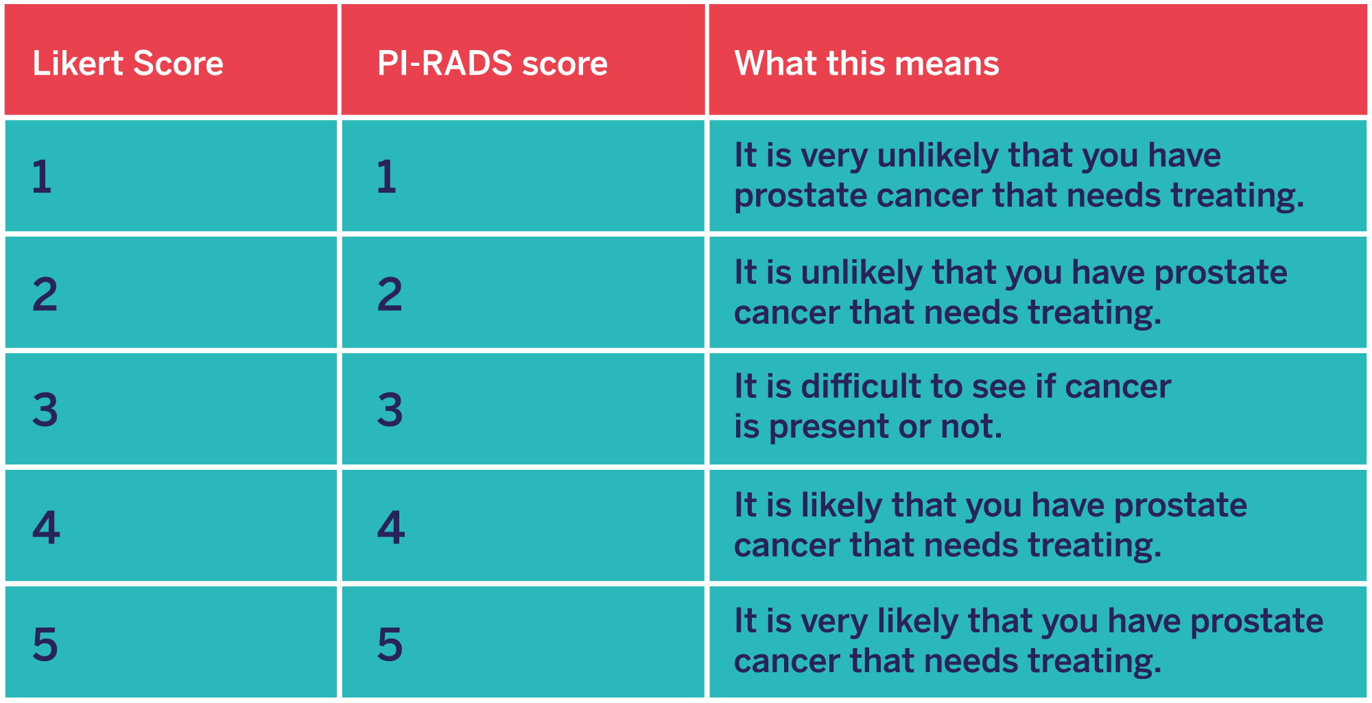
  • A radiologist will look at your scans and give them a score. 
  • You may hear your doctor talk about the Likert score or the PI-RADS score. 
  • These go from one-to-five and tell you how likely it is you have prostate cancer. This will help your doctor decide what to do next. 
Pi-Rads score

About this information

  • This information was published in March 2023. We will revise it in March 2024.
  • References and bibliography available on request.
  • If you want to reproduce this content, please see our Reproducing Our Content page.

Share your story with others

By sharing your story you can help others. Share what went well, what didn't and everything in between. Your experience is valuable. Help make the prostate cancer community stronger.

Share story